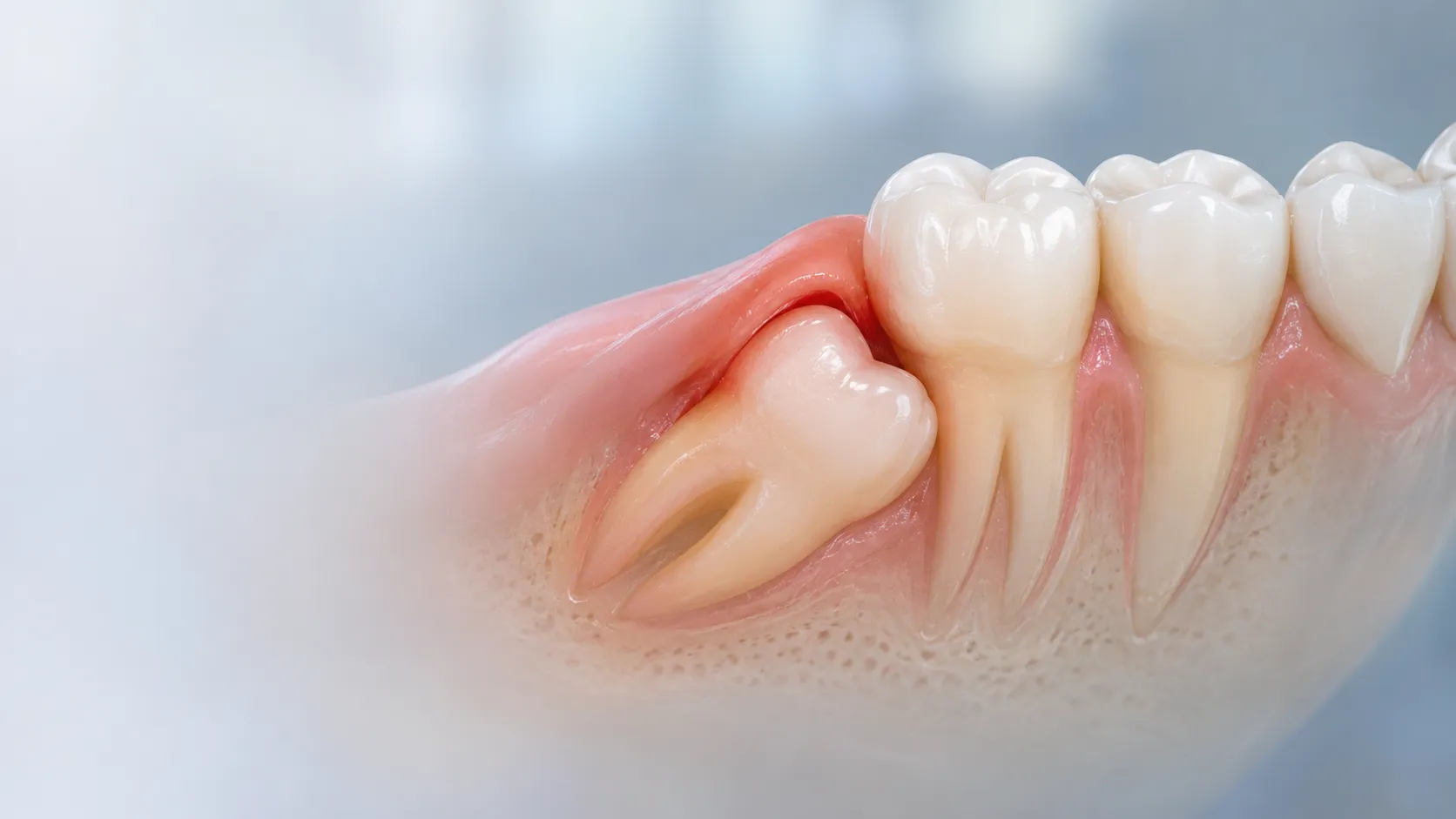
When an oral surgeon reviews your panoramic digital X-ray or 3D scan, one of the first things they evaluate is the exact direction or “angle of impaction” of your third molars. An impacted tooth simply means a tooth that has failed to erupt into its normal functional position within the dental arch because of structural blockage or a lack of jaw space.
However, not all impactions carry the same level of surgical complexity or risk. Understanding the structural differences between vertical, mesial, distal, and horizontal impactions can help you make informed decisions about your dental care.
Body
- Understanding the Four Primary Angles of Impaction
- The orientation of a third molar plays a direct role in how it impacts your jawbone, nerve pathways, and neighboring teeth:
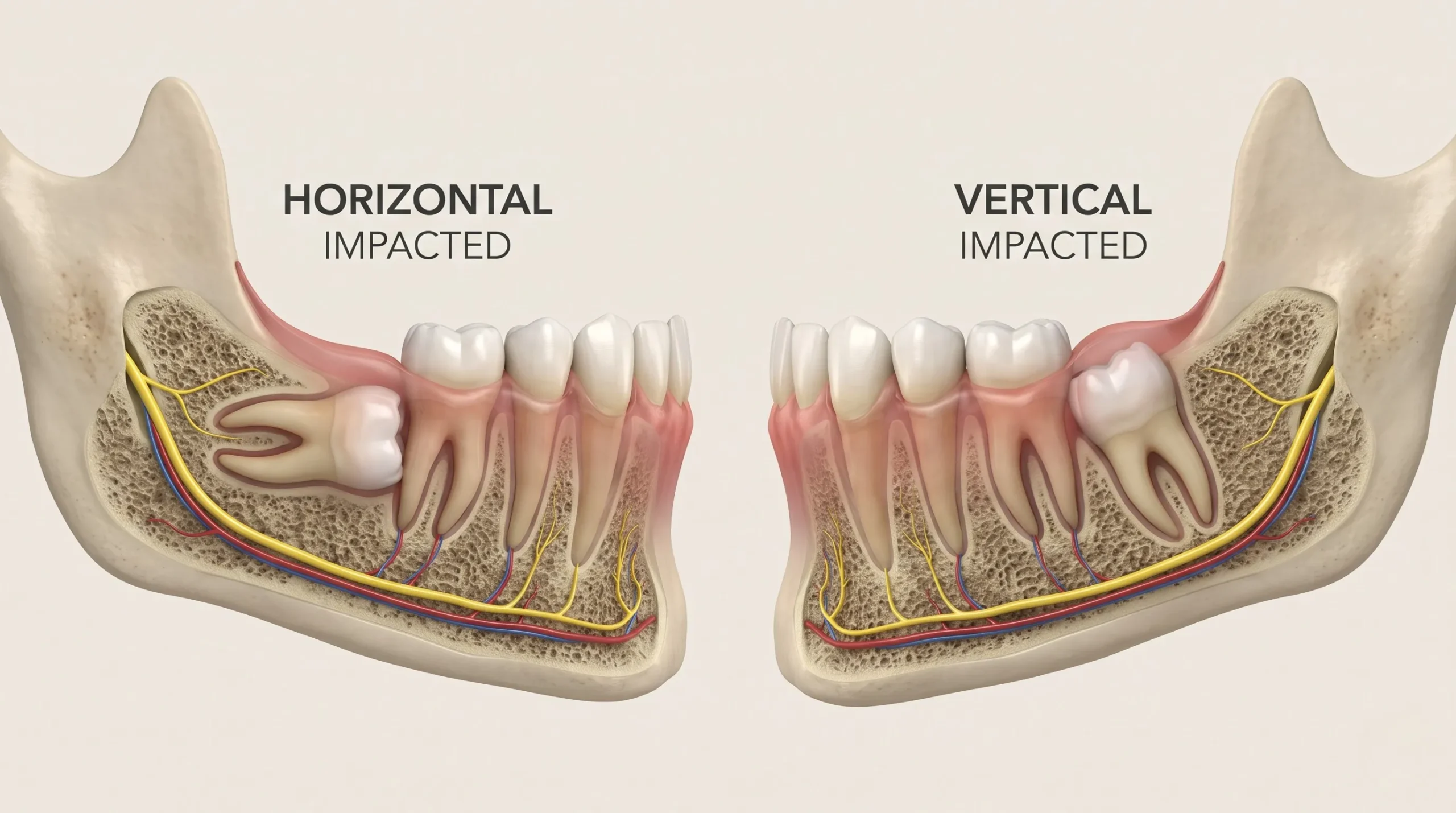
Vertical Impaction (Straight Up-and-Down): The tooth is oriented in the correct vertical position for normal eruption.
However, it remains trapped beneath thick gum tissue or jawbone because the jaw arch lacks the physical length to accommodate it.
Mesial Impaction (Angled Forward toward the Front of the Mouth): This is the most frequently diagnosed type of impaction.
The crown of the wisdom tooth tilts forward at a 45-degree angle, pushing directly into the upper back corner of the second molar.
Distal Impaction (Angled Backward toward the Throat): The tooth angles backward toward the rear of the jaw bone.
It remains buried deep within the ascending ramus of the mandible, often requiring precise bone access for removal.
Horizontal Impaction (Lying Sideways at a 90-Degree Angle): The tooth is lying completely on its side beneath the gums.
The top enamel crown points straight into the roots of your adjacent healthy second molar, making this the most complex type of impaction.
- Why Horizontal Impactions Demand Timely Treatment
- Leaving a sideways horizontal impaction untreated creates severe long-term complications across your jaw:
Destruction of Second Molar Roots (External Root Resorption): As the sideways tooth attempts to erupt, it exerts continuous physical pressure on the root system of the second molar, slowly dissolving its healthy bone and tooth structure.
Uncleanable Deep Cavity Pockets: The junction where a sideways wisdom tooth hits an adjacent molar creates an unreachable food trap. This leads to deep tooth decay below the gumline on both teeth.
Cyst Formation and Jaw Expansion: Horizontal teeth often remain completely encased in the jawbone. Over time, the fluid-filled sac surrounding the tooth crown can expand into a dentigerous cyst, eroding jawbone matrix.
- How Modern 3D CBCT Scans Make Extractions Predictable
- Exact Nerve Mapping: A 3D CBCT scan creates a millimeter-precise, three-dimensional reconstruction of your jaw, allowing oral surgeons to map the exact distance between the wisdom tooth roots and the inferior alveolar nerve trunk.
Micro-Surgical Sectioning: Rather than forcefully pulling a horizontal tooth, oral surgeons use ultrasonic piezosurgery instruments to section the tooth into tiny, effortless pieces. Each section is gently lifted out without applying force to surrounding jawbone.
Conclusion
The specific angle of your wisdom tooth determines its surgical pathway and long-term impact on your mouth. Catching horizontal and angular impactions early through 3D imaging protects your adjacent healthy molars from irreversible structural damage.
Curious about the orientation of your wisdom teeth? Schedule a low-radiation 3D CBCT diagnostic assessment at Pearl Dental Kochi to plan your care with precision.